Chiari malformation
Neuroinstitute: Innovative technology for safe surgery
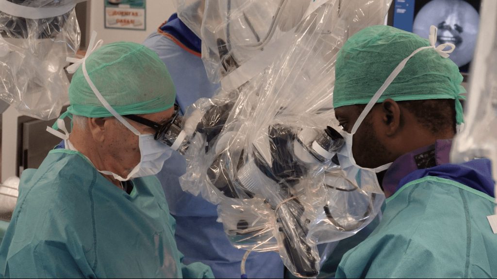
Surgical treatment of chiari malformation
Explained by Dr. Bartolomé Oliver & Dr. Nnamdi Elenwoke
Chiari syndrome is a congenital malformation at the base of the skull, where brain tissue extends into the spinal canal. This can cause a variety of debilitating symptoms.
Surgical treatment is recommended when symptoms are severe or progressive .
Depending on the clinical manifestations, we use different types of surgical intervention.
At Neuroinstitute we have more than 50 years of experience in surgical treatment of Chiari syndrome. Our experience and dedication ensure high quality care for our patients.
Diagnosis
Chiari syndrome can occur in patients of all ages, but is more common in young women. It occurs due to abnormalities in the development of the skull and spinal cord.
Symptoms of Chiari syndrome can range from severe headaches to coordination problems and weakness. These symptoms occur due to compression of brain tissue and impaired flow of cerebrospinal fluid.
Chiari syndrome can cause serious complications, such as hydrocephalus, loss of neurological function, and even paralysis. It is dangerous because it can significantly affect quality of life and require surgical intervention
-
Chiari type 0
Type 0 is very rare. In this type, there are little to no parts of your cerebellum in the hole at the base of your skull (foramen magnum) but there is crowding at that level.
Symptoms occur because of an abnormal flow of cerebrospinal fluid near the base of your skull.
-
Chiari type I
In this subtype, brain tissue, primarily the cerebellum, moves downward through the foramen occipital.
It is the most common type and is usually diagnosed in adolescence or adulthood.
-
Chiari type II
This subtype is commonly associated with spina bifida, a congenital anomaly of the spinal cord.
In Chiari type II, a descent of the cerebellum and brain stem into the spinal canal is seen.
-
Chiari type III
It is the rarest type and is characterized by the partial or complete absence of the cerebellum.
This can cause serious neurological problems and is associated with more complex brain malformations.
-
Chiari type IV
This is a rarer and more complex form of the disease, in which brain tissue is wedged into the skull due to developmental defects. Patients with type IV Chiari malformation suffer from severe neurological complications.
Diagnostic methods
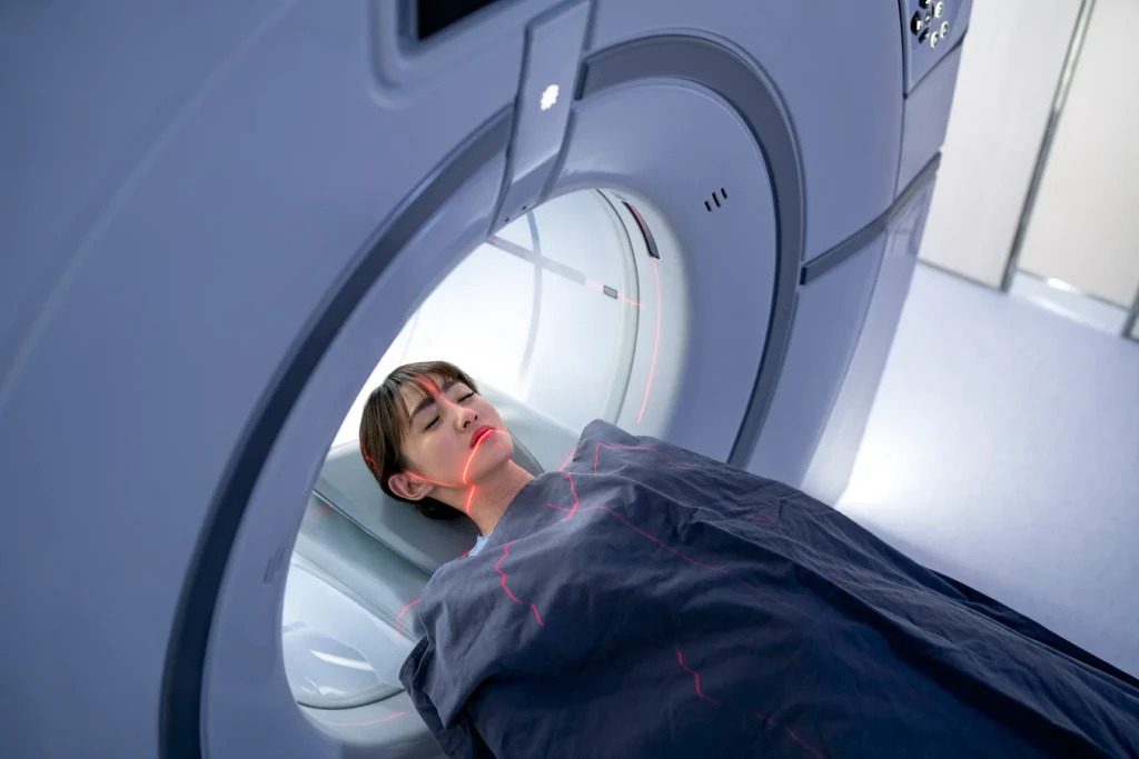
Magnetic Resonance Imaging (MRI) of the brain and spine
MRI is the most commonly used diagnostic test to evaluate Chiari malformation. It allows detailed visualization of the anatomy of the brain, cerebellum and spinal cord, identifying any abnormalities in the structure and determining whether brain tissue has moved into the spinal canal.
Computed Tomography (CT) of the brain
Computed tomography can be used as an alternative to MRI in cases where MRI is not possible, although it is not as effective in visualizing soft tissues such as the brain and spinal cord.
X-ray of the cervical spine
Can be helpful in evaluating the alignment of the vertebrae and detecting possible abnormalities that may contribute to Chiari malformation.
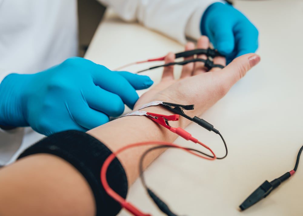
Electromyography (EMG) and Nerve Conduction Studies
These tests may be performed to evaluate peripheral nerve function and detect possible signs of nerve pathology associated with Chiari malformation.
Leading doctors
-

35+ years of experience
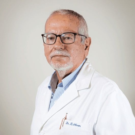
Dr. Bartolomé Oliver
CEO and Medical Director of Neurosurgery Institute Leading neurosurgeon
Dr. Bartolome Oliver Abadal is a founder of the Neuroinstitute based at Teknon Medical Centre, Barcelona.
Graduated from the Faculty of Medicine at the University of Barcelona in 1977. He completed an internship at the National Department of Neurosurgery in Madrid.
He continued his internship at the Hospital de Sant Pau University Hospital in Barcelona, and completed internships in hospitals in Stockholm (Sweden), Zurich (Switzerland), Montreal (Canada), St. Louis (USA) and Pittsburgh (USA).
The author of more than 150 scientific papers and 350 international reports.
Full cvSpecialization
Complex neurosurgery of skull and spine
Chiari malformation surgery
Minimally invasive endoscopic neurosurgery of skull base
Craniocervical Instability
Hydrocephalus and syringomyelia
Vascular neurosurgery
-
17+ years of experience
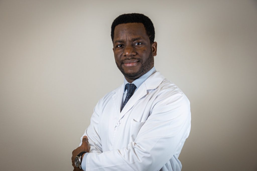
Dr. Nnamdi Elenwoke
Neurosurgeon
Dr. Nnamdi Elenwoke is a renowned neurosurgery specialist with extensive experience in minimally invasive surgery and robotic surgery.
After completing his medical training in 2007, Dr. Elenwoke carried out several scientific studies. He completed the Neurosurgery residency at the Miguel Servet Hospital in Zaragoza and trained in endoscopic skull base technique sat Emory University, Atlanta USA.
Continuing to expand and improve his skills, Dr.Elenwoke gained practice in the best neurosurgery centers, such as the Hospital for Neurology and Neurosurgery London (UK), University Hospital Araba de Santiago (Spain) and joined the prestigious team of Neuroinstitute at the Teknon Medical Centre and Hospital of Barcelona.
Full cvSpecialization
Chiari malformation surgery
Hydrocephalus and syringomyelia
Craniocervical instability
Minimally invasive surgery of skull and spine
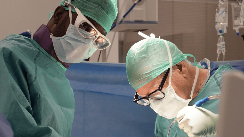
Surgical treatment
There are different types of surgery for Chiari syndrome, such as craniocervical decompression and cervical laminectomy. Each has its own benefits and risks.
At our institute, we use state-of-the-art intraoperative navigation systems, such as Medtronic's StealthStation™, which allows for optimal surgical precision and better visualization of anatomical structures during surgery.
Meticulous surgical protocols are followed and specialized post-operative care is provided to minimize risks and promote successful recovery
-
Craniocervical
decompressionSurgical technique:
The craniocervical area is accessed through an incision in the back of the head and neck.
Part of the bone of the skull and spine is removed to relieve compression of the brain and restore the normal flow of cerebrospinal fluid.
Results:
Significant improvement of neurological symptoms, pain relief and functional recovery in many patients.
Improves quality of life, prevents progression of symptoms and reduces the risk of serious complications.
-
Cervical laminectomy
Surgical technique:
An incision is made in the back of the neck to access the cervical region.
The vertebral arch is removed to decompress the spinal canal and relieve compression of the nervous tissue.Results:
Improvement of neurological symptoms associated with spinal cord compression and restoration of neurological function.
Reduction of pain, improvement of mobility and prevention of serious neurological complications.
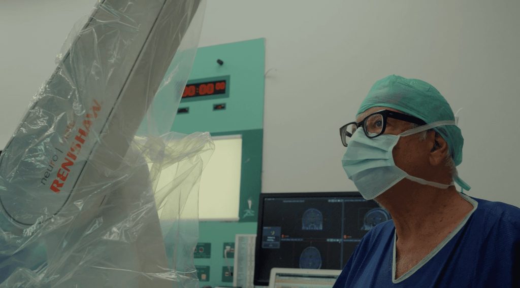
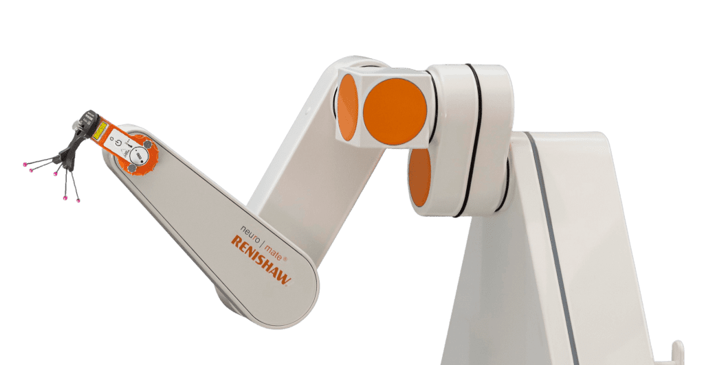
Innovative technology
Robotic Neurosurgery System
Neuromate Renishaw
The Neuromate™ robot allows us to perform complex neurosurgical procedures with sub-millimeter precision.
Based on the results of CT and MRI, the robot is programmed individually for the anatomy of each person.
It guarantees maximal safety during the surgery and avoids surgical complications.
-
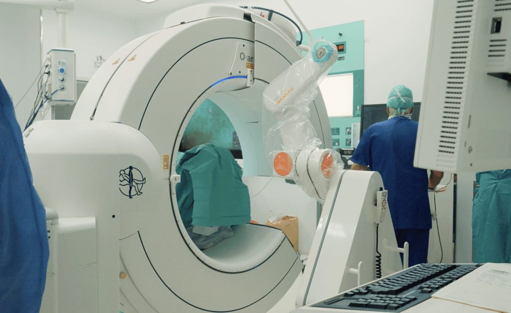
O-ARM SURGICAL IMAGING
The O-arm™ system is an intraoperative 2D/3D imaging system that is designed for achieving radiological images during the surgery.
The O-arm system’s high quality, versatile imaging provides the anatomical information for clinical decision making right in the surgery room.
Along with StealthStation navigation, the O-arm system provides enhanced 3D visibility and surgical feedback.
-
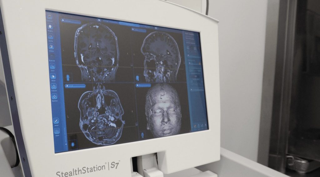
StealthStation S7 Neuronavigation
The StealthStation™ S7 surgical navigation system enables to precisely track the location of surgical instruments throughout a procedure.
It combines optical and electroneuromyography monitoring to avoid damage of nervous tissue and exclude complications such parálisis or function loss.
Support on each step
of your treatment
-
Request a free consultation
with medical assistantOur medical assistant will contact you to find out how we can help you.
They will help prepare your case for a consultation with the doctors. -
Prepare your medical
reports and scansSend your medical reports, CT & MRI scans to your assistant and he will review them and prepare them for the doctors. So no detail is missed.
-
Get an appointment
with the doctorsConsult with doctors online without leaving home. Your assistant will arrange a video call with the Doctors to discuss your treatment plan and receive an official medical report.
-
Prepare for your trip
and receive treatmentOnce you have made a decision about treatment, our medical assistant will prepare a budget and help organise your visit. The clinic provides visa support.
-
Treatment
& hospital stayHospitalisation in a comfortable individual room on the ward with Barcelona city views.
Daily control visits by doctors, a nutritionist, and round-the-clock medical supervision. -
Follow-up
after treatmentWe stay in touch with you even after your return home and control the result of treatment.
You can always ask questions to your doctor
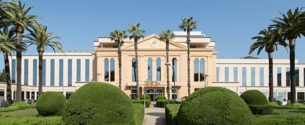
Hospital
Neuroinstitute operates at the clinical base of the Teknon Medical Centre, one of the largest multidisciplinary hospitals in Spain, which allows the patient to receive any necessary medical care and provide the necessary care and supervision in the postoperative period.
The hospital regularly undergoes international certification and has been certified by Joint Commission International seven times, which guarantees the high quality and safety of medical services.
Teknon Medical Centre — the best international hospital
MTJ: International Medical Travel Jornal

What our patients have to say
For doctors
Neuroinstitute collaborates with doctors and clinics around the world to share knowledge and experience in the fight against neurosurgical pathology.
If you are a medical professional interested in cooperation, please fill out the form and we will contact you as soon as possible.